
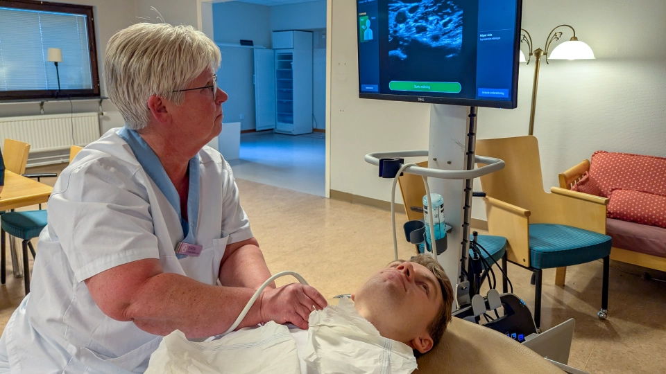
Researchers and hospital engineers from the North Sweden ecosystem are developing a new way to detect atherosclerosis in primary care. By making vascular ultrasound more accessible through AI support, the technology could help people understand their personal risk, support healthier lifestyles and contribute to more equitable preventive care, particularly in rural areas.
Residents of Västerbotten are invited to a health examination and counselling session when they turn 40, 50 or 60. The Västerbotten Intervention Programe helps identify people at increased risk of cardiovascular disease and supports changes in diet, physical activity and other lifestyle factors. Preventive medication may also be considered when needed.
Research linked to the programe shows that cardiovascular risk is generally higher in rural areas, particularly among men. The difference is mainly associated with lifestyle factors rather than access to care. An accessible tool that makes risk visible could therefore be especially valuable in smaller communities far from hospital-based specialist services.
“If you understand your risks and hereditary factors, and can also see what your carotid arteries look like, it may become easier to consider medication and change habits such as diet and physical activity.” Greta Svahn, nurse at Malå cottage hospital
The Primviza research project builds on Vipviza, a study conducted at Umeå University since 2013. Vipviza investigates whether ultrasound images of the carotid arteries, combined with health counselling, can help people understand their cardiovascular risk. Primviza is developing methods that could bring this type of imaging from specialist hospital care into primary care.
Ultrasound images can reveal fatty deposits in the carotid arteries, a sign of atherosclerosis and increased cardiovascular risk. The progression of atherosclerosis can be slowed and may even be reduced through healthier lifestyle choices. Region Västerbotten’s Medical Technology Research and Development unit is also working on visual presentations that make the results easy for both patients and healthcare professionals to understand.
Gel is applied to the neck to ensure good contact with the skin. The examiner moves a handheld ultrasound probe while on-screen guidance helps identify the correct areas of the carotid arteries. The AI technology has been trained on a large number of examples to recognise normal and altered blood vessels. The results are intended to be presented as clear, accessible images.
A prototype has been tested by staff at Malå cottage hospital, where distances to hospital care are long. The trial showed that the technology is moving towards being manageable for primary care staff, although further research and training are still needed before implementation can be considered.
“It would take some practice, but it would be worth it. I think it becomes easier for people to make a change when they can see the images this clearly.” Maria Larsson, assistant nurse at Malå Health Centre
If the technology becomes available in smaller communities, it could strengthen the Västerbotten Intervention Programme and contribute to more equitable preventive care. No date has yet been set for implementation, but the researchers see the Malå test as an important step towards a practical solution for primary care.
Primviza is led by Patrik Wennberg, professor at Umeå University, in collaboration with primary care and Medical Technology Research and Development at Region Västerbotten. Researchers and development staff involved in the Malå test included Albin Dahlin Almevall, Jakob Marklund, Karolina Jonzén and Christer Grönlund, together with staff from Malå Health Centre.